


Medical Hair Encyclopedia
Dermatological pathologies and evidence-based clinical guidelines at international standards.
CLINICAL GUIDELINE & PATHOLOGY
Seborrheic Dermatitis (Scalp Eczema) – Diagnosis and Treatment Guide
📌 Patient Information Section: What Does This Condition Mean?
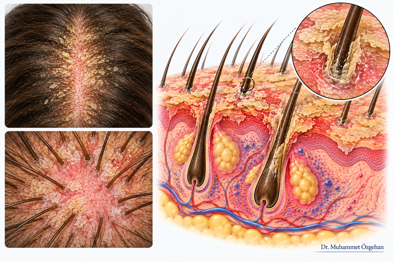
Seborrheic dermatitis is a common type of eczema with a chronic course that can be controlled, appearing on the scalp and in areas with a high concentration of sebaceous glands (between the eyebrows, sides of the nose, behind the ears). It is not contagious.
- What Causes It?: The primary triggers are the overgrowth of a yeast fungus called Malassezia, which naturally resides on the scalp, the skin's sensitive immune response to this fungus, and the quality of sebum (skin oil) secretion. Stress, fatigue, seasonal changes, and cold weather attacks can exacerbate it.
- What Are Its Symptoms?: Dandruff on the scalp, mild or severe redness, oily and yellowish scales, and an occasionally bothersome itching sensation.
- What Should Be Done?: Using random shampoos or irritating the skin by thinking "it will pass" can worsen the condition. The disease can be kept under control with dermatologically approved medical shampoos and appropriate care products.
🩺 Clinical & Medical Guide for Physicians
- Etiology and Pathophysiology: Increased lipid metabolites caused by Malassezia yeast species, keratinocyte proliferation, and accompanying mild-to-moderate perivascular inflammation.
- Diagnostic Criteria and Clinical Findings: Widespread erythema on the scalp and border areas, with slightly oily, white-yellowish scales. The appearance of a "seborrheic crown" extending along the frontal hairline.
- Dermoscopy (Trichoscopy) Findings: Zigzagging fine telangiectasias, diffuse yellowish-pink erythema in the interfollicular area, and oily dandruff islands (follicular plugs) obstructing hair ostia (distinguished from psoriasis by the absence of follicular plugs).
- Histopathological Diagnosis (Punch Biopsy):
- Biopsy Indication and Technique: In atypical, resistant cases and those where differential diagnosis from psoriasis is difficult, a 4 mm punch biopsy should be taken from the lesion. Examination is performed with vertical sections.
- Pathological Findings: Superficial perivascular lymphocytic infiltrate, mild acanthosis, mild spongiosis (edema) in follicular ostia, and "fungal nests" (parakeratotic plugs) containing neutrophils in the infundibulum.
- Current Treatment Protocols:
- Topical Antifungals and Keratolytics: Medical shampoos containing ketoconazole, ciclopirox olamine, or zinc pyrithione. Removal of scales with shampoos containing salicylic acid or selenium sulfide.
- Topical Anti-inflammatories: Short-term use of weak/moderate potent topical corticosteroid lotions or calcineurin inhibitors during flare-ups. Additionally, expertise and treatment approaches can be reviewed for clinical and surgical evaluations.
Scientific References and Bibliography
- Clark GW, Pope SM, Jaboori KA. Diagnosis and treatment of seborrheic dermatitis. Am Fam Physician. 2015 Feb 1;91(3):185-90. PMID: 25822272. PubMed Link
- Borda LJ, Perper M, Keri JE. Treatment of seborrheic dermatitis: a comprehensive review. J Dermatolog Treat. 2019 Mar;30(2):158-169. doi: 10.1080/09546634.2018.1473554. Epub 2018 May 24. PMID: 29737895. PubMed Link
- LeFevre NM, Braudis K, Feigenbaum LS. Seborrheic Dermatitis: Diagnosis and Treatment. Am Fam Physician. 2025 Aug;112(2):166-173. PMID: 40834373. PubMed Link