


Medical Hair Encyclopedia
Dermatological pathologies and evidence-based clinical guidelines at international standards.
CLINICAL GUIDELINE & PATHOLOGY
Hair Involvement in Systemic Infections (Secondary Syphilis and "Moth-eaten" Alopecia) – Diagnosis and Treatment Guide
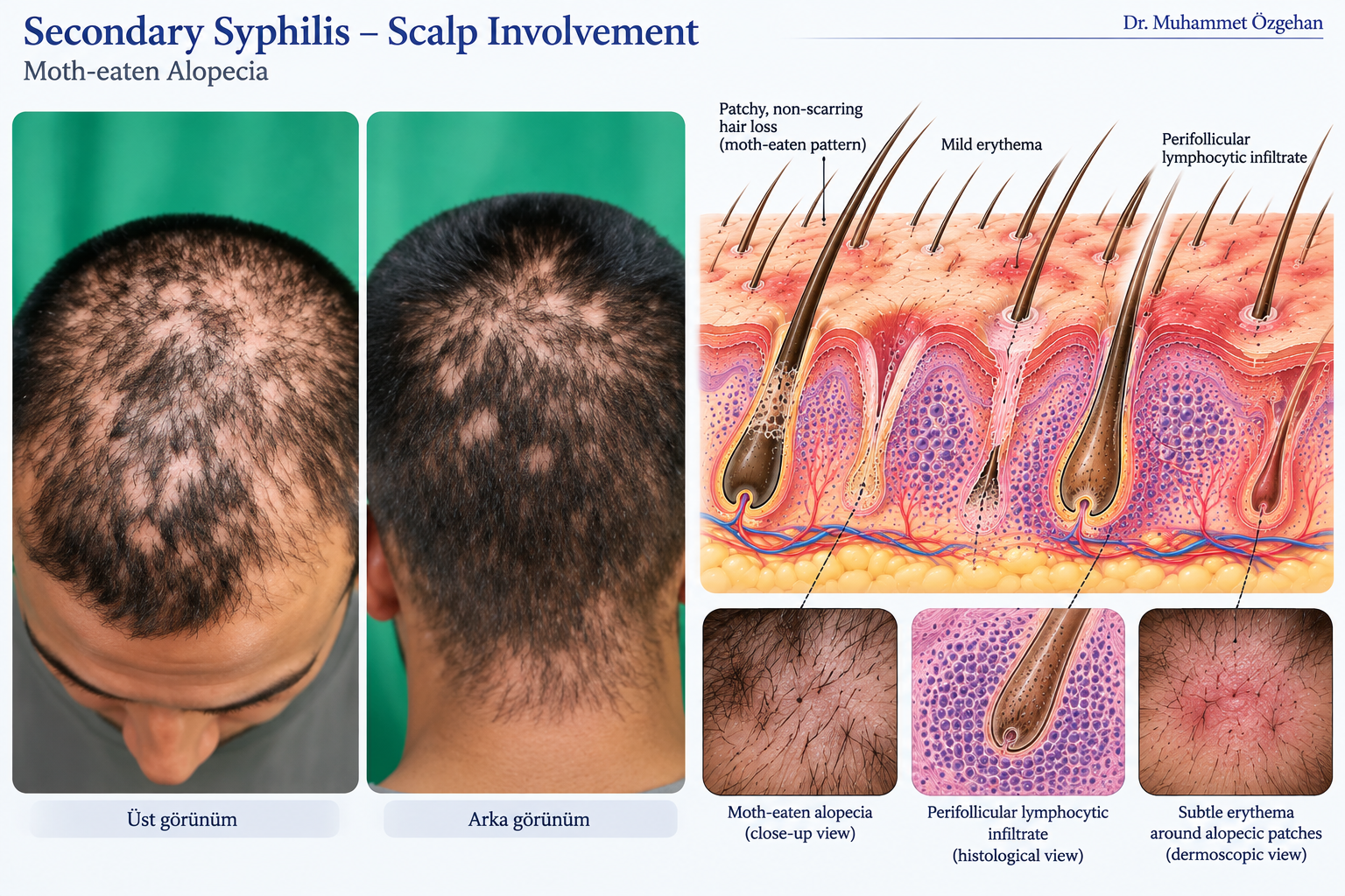
📌 Patient Information Section: What Does This Condition Mean?
In the second stage of syphilis, a sexually transmitted systemic infection, a very characteristic pattern of hair loss can emerge on the scalp, in addition to general body symptoms.
- What is Moth-Eaten Alopecia?: On the scalp, particularly in the occipital (back of the head) and temporal (temple) regions, multiple small, scattered patches of hair loss, coin-sized and without sharp borders, appear. The appearance resembles fabric eaten by moths.
- Other Findings: Pale red rashes on the palms and soles, widespread lymph node swelling, and oral lesions may accompany this condition.
- What to Do?: This presentation is indicative of a systemic disease. Local hair treatments are ineffective; serological tests should be performed early, and penicillin treatment initiated under expert supervision.
🩺 Clinical & Medical Guide for Physicians
- Etiology and Pathophysiology: Systemic dissemination of the spirochete bacterium Treponema pallidum. Endarteritis in the vessels surrounding hair follicles, leading to ischemic/inflammatory hair loss (non-scarring alopecia).
- Diagnostic Criteria and Clinical Findings: "Moth-eaten" alopecia, seen in approximately 4-7% of secondary syphilis patients. Hair loss in the lateral part of the eyebrows may also be present. Serological positivity for VDRL/RPR and TPHA/TPPA.
- Dermoscopy (Trichoscopy) Findings: Absence of black dots (broken hair shafts), empty follicular ostia, preserved interfollicular epidermis, and telangiectatic vascular patterns on a yellowish-pink background.
- Histopathological Diagnosis (Punch Biopsy):
- Biopsy Indication: A 4 mm punch biopsy is taken from the lesion when atypical cases are suspected and confused with alopecia areata.
- Pathology: Dense infiltrates rich in plasmocytes in the perivascular and perifollicular areas (plasmocyte infiltration is highly typical for syphilis) and findings of endarteritis.
- Current Treatment Protocols:
- Systemic Treatment: Benzathine Penicillin G 2.4 million units IM single dose (in the secondary stage). Doxycycline or Azithromycin for those with penicillin allergy. With treatment, hair spontaneously and completely regrows within a few months (it is non-scarring). Additionally, specialized and treatment approaches can be reviewed for clinical and surgical evaluations.
Scientific References and Bibliography
- Dai S, Wang H, Lin Z. Moth-eaten alopecia in secondary syphilis. Int J Infect Dis. 2019 May;82:6. doi: 10.1016/j.ijid.2019.02.023. Epub 2019 Feb 23. PMID: 30807867. PubMed Link
- Saraswat N, Tripathy DM, Kumar S, Ranjan E, Madakshira MG, Goel S. Malignant secondary syphilis presenting with rupioid lesions and moth-eaten alopecia. Indian J Sex Transm Dis AIDS. 2024 Jan-Jun;45(1):82-83. doi: 10.4103/ijstd.ijstd_73_23. Epub 2024 Jun 6. PMID: 38989088; PMCID: PMC11233054. PubMed Link
- Jordaan HF, Louw M. The moth-eaten alopecia of secondary syphilis. A histopathological study of 12 patients. Am J Dermatopathol. 1995 Apr;17(2):158-62. doi: 10.1097/00000372-199504000-00008. PMID: 8600781. PubMed Link