


Medical Hair Encyclopedia
Dermatological pathologies and evidence-based clinical guidelines at international standards.
CLINICAL GUIDELINE & PATHOLOGY
Tinea Capitis (Scalp Fungal Infection) – Diagnosis and Treatment Guide
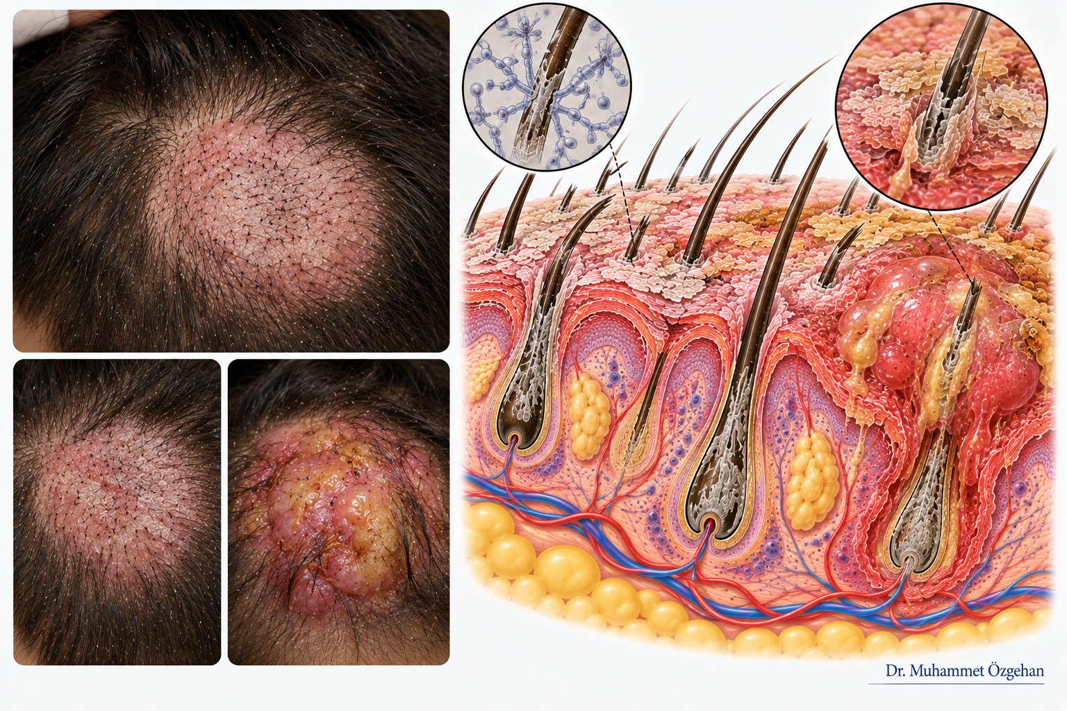
📌 Patient Information Section: What Does This Condition Mean?
Tinea capitis is a scalp disease caused by a contagious fungal infection, often incorrectly referred to by the public as "ringworm" (confused with Alopecia areata).
- Who is Affected?: It is most commonly seen in school-age children; however, it can also be transmitted to adults. It spreads very easily in schools, nurseries, or through the shared use of combs, towels, and hats. Transmission can also occur from cats, dogs, or farm animals (zoophilic species).
- What are the Symptoms?: It may start as slightly scaly, mildly erythematous (reddened) circular areas on the scalp. Over time, the hair in these areas weakens and breaks off at the skin level (black dot appearance). In some types, it can form inflammatory, soft, raised swellings that ooze pus upon pressure (honeycomb appearance), known as "Kerion."
- What Should be Done?: It never resolves with homemade oils or dandruff shampoos. Because it is contagious and can lead to permanent hair loss if left untreated, a specialist physician should be consulted immediately, and family members should also be screened.
🩺 Clinical & Medical Guide for Physicians
- Etiology and Pathophysiology: Dermatophyte species (Microsporum canis, Trichophyton mentagrophytes, Trichophyton tonsurans, etc.) are the causative agents. Fungal spores invade the stratum corneum and the hair shaft (with endothrix or ectothrix localization).
- Diagnostic Criteria and Clinical Findings: Scaly, grayish patch areas where hair breaks off close to the skin. In severe cases, Kerion Celsi (developing as a result of secondary bacterial infection and excessive immune response), characterized by deep, painful, purulent abscess formation, and cervical lymphadenopathy.
- Dermoscopy (Trichoscopy) Findings: "Comma hairs" formed by the breaking of the hair shaft at the skin level, "corkscrew hairs," "zigzag hairs," and gray-white scale accumulations in the follicular ostia.
- Histopathological Diagnosis and Microbiological Confirmation:
- Diagnostic Methods: Obtaining a scalp scraping or epilation sample for culture is the gold standard. Direct visualization of fungal spores (arthroconidia) within or around the hair shaft in a 10% KOH (Potassium Hydroxide) preparation.
- Biopsy and Pathology: A 4 mm punch biopsy is taken in cases of suspected Kerion or atypical cases. Fungal hyphae and spores within the hair shaft are clearly observed with PAS (Periodic acid–Schiff) staining. Dense neutrophilic and granulomatous inflammation is seen in the dermis.
- Current Treatment Protocols:
- Systemic Treatment is Essential: Topical shampoos alone are insufficient. Systemic antifungal treatments (Griseofulvin, Terbinafine, Itraconazole, or Fluconazole) should be administered for at least 6-8 weeks, calculated according to weight in children.
- Kerion Management: In the presence of Kerion, antibiotic support for secondary bacterial infection and short-term systemic corticosteroids (to prevent inflammation and permanent scarring) may be added. Family screening and cessation of shared item use are mandatory. Additionally, expertise and treatment approaches can be reviewed for clinical and surgical evaluations.
Scientific References and Bibliography
- Gupta AK, Friedlander SF, Simkovich AJ. Tinea capitis: An update. Pediatr Dermatol. 2022 Mar;39(2):167-172. doi: 10.1111/pde.14925. Epub 2022 Jan 24. PMID: 35075666. PubMed Link
- Leung AKC, Hon KL, Leong KF, Barankin B, Lam JM. Tinea Capitis: An Updated Review. Recent Pat Inflamm Allergy Drug Discov. 2020;14(1):58-68. doi: 10.2174/1872213X14666200106145624. PMID: 31906842. PubMed Link
- Gupta AK, Polla Ravi S, Wang T, Faour S, Bamimore MA, Heath CR, Friedlander SF. An update on tinea capitis in children. Pediatr Dermatol. 2024 Nov-Dec;41(6):1030-1039. doi: 10.1111/pde.15708. Epub 2024 Aug 7. PMID: 39113245. PubMed Link